Monday, January 30, 2023

Long-Covid Diagnosis and treatment

 Long-Covid Diagnosis and Treatment


Long COVID-19 is estimated to affect approximately 1.4 million Canadians. In clinic, I happened to see a suspected case. Here I will discuss the diagnosis and treatment of Long COVID syndrome. 

Diagnosis: 

The current case definition put forth by the world health organization describes long-COVID as: symptoms that linger beyond 3 months of a probable or confirmed SARS-CoV-2 infection, which last at least 2 months and cannot be explained by an alternative diagnosis. 

However, there are not yet specific diagnostic tests that have a high enough negative or positive predictive value that can rule in or rule out disease. ie, this is currently a clinical diagnosis.
Interestingly, confirmed SARS-CoV-2 infection need not be presented via PCR or RAT. Instead a likely case ie. high-risk exposure with symptoms qualifies in this case definition. 

Perhaps the most important part of this case definition is that alternative diagnoses must be ruled out. Ie, if the one of the patient's symptoms include a headache, a primary headache disorder such as migraines or a secondary headache disorder such as GCA must be ruled out.

Treatment: 

While there is not specific or definitive treatment for Long-COVID, features of the syndrome are treated symptomatically.

Fatigue and Post-Exertional malaise

This is a common symptom of long-covid and recommended treatment includes: 
a structured and symptom-guided return to activity program, tailored to their severity of fatigue. The 4 Ps (pacing; prioritizing which activities need to get done on specific days and which activities can be postponed; positioning to modify activities to make them easier to perform [e.g., while sitting]; and planning)

Mental health complications of long COVID

Common mental health complications of long COVID include anxiety depression and and post-traumatic stress disorder. The recommended treatment of these conditions is guideline based medical therapy. Referral to a psychiatrist can also be considered. 

Dyspnea

In people with mild dyspnea, pursed lip or deep breathing exercises may improve symptoms. While persistent hypoxemia is rare, it should prompt Respirology referral to rule out lung pathologies such as an organizing pneumonia

Sleep disturbances
Patients should receive counselling on sleep hygiene, relaxation techniques and stimulus control. Cognitive behavioural therapy is an option for treating sleep disturbances. Alternatively, medical therapy may also include management with: eszopiclone, zolpidem or doxepin

Palpitations/Tachycardia
Options for treating inappropriate sinus tachycardia include: behavioural modifications, oral fluids, salt, compression stockings, β-blockers, ivabradine and midodrine

References: 


-AM-


Friday, January 27, 2023

Revised Cardiac Risk index (RCRI)

RCRI is an index to evaluate major cardiac complications for noncardiac surgery. There are 6 risk predictors of RCRI:

  1. High risk surgery (e.g., vascular, intrathoracic) (Odds Ratio 2.6)
  2. CAD (e.g., hx of MI, PCI, CABG, Angina, nitrate use, ECG with pathological Q wave, or positive stress test) (Odds Ratio 3.8)
  3. History of CHF, pulmonary edema, bilateral rales (Odds Ratio 4.3)
  4. History of cerebrovascular disease (e.g., stroke or TIA) (Odds Ratio 3)
  5. Diabetes mellitus treated with insulin (Odds Ratio 1)
  6. Preop Cr > 176.8 µmol/L (Odds Ratio 0.9)
Note that those Odds Ratios above were calculated using multivariable logistic regressions rather than univariate logistic regression. 


The original RCRI article showed event rate as below:

Rate of cardiac death, MI, pulm edema, complete heart block, or VF/cardiac arrest

Number of RCRI Risk factors

Rates of event

0

0.5%

1

1%

2

5%

3

10%


The rates of major cardiac complications were later reassessed in 2017 using four data from four prospective studies and one retrospective studies:

Rate of myocardial infarction, cardiac arrest, or death at 30 days after noncardiac surgery

Number of RCRI Risk factors

Rates of event

0

4%

1

6%

2

10%

3

15%


The discrepancies were potentially caused by the test method and enrollment criteria. The original study used CK-MB while recent studies used troponin which were more sensitive. Additionally, the original study excluded emergency surgery patients while recent studies included patients who received emergency surgeries. 


Reference:

Canadian Cardiovascular Society Guidelines on Perioperative Cardiac Risk Assessment and Management for Patients Who Undergo Noncardiac Surgery - PubMed (nih.gov)

Derivation and Prospective Validation of a Simple Index for Prediction of Cardiac Risk of Major Noncardiac Surgery | Circulation (ahajournals.org)

Revised Cardiac Risk Index for Pre-Operative Risk - MDCalc


-YZ-

Primary hyperparathyroidism

 Hyperparathyroidism is defined as a disorder which involves over secretion of parathyroid hormone (PTH). Given the etiology, physicians often classify it as primary, secondary, and tertiary causes. 

Primary hyperparathyroidism most commonly caused by parathyroid adenoma or hyperplasia. MEN type 1 and 2A are potential risk factors this presentation. Usually, patient was asymptomatic or have mild nonspecific symptoms including fatigue and constipation. Other potential symptoms including abdominal pain, bone pain, neuropsychiatric symptoms, and symptoms from nephrolithiasis. 

Diagnostic findings of primary hyperparathyroidism include elevated or inappropriately normal PTH in the context of hypercalcemia. Other conditions with similar lab findings include Familial Hypocalciuric Hypercalcemia which could commonly be differentiated by testing the urinary calcium clearance to creatinine clearance ratio (UCCR). Besides FHH, Tertiary hyperparathyroidism (pts with primary hyperparathyroidism often have low phosphate but patients with tertiary hyperparathyroidism often have high phosphate level) and rarely ectopic PTH production are also in this PTH dependent hypercalcemia category. 

In general, asymptomatic primary hyperparathyroidism patients can be managed conservatively initially. Common measures include encourage oral hydration, encourage physical activity, and avoid high calcium diet as well as certain medications (e.g., thiazide, lithium). 

For primary hyperparathyroidism patients, surgery can be curative. Indications for parathyroidectomy include:

  1. Age < 50
  2. Symptomatic hypercalcemia
  3. Complications: osteoporosis, previous asymptomatic vertebral fracture, nephrolithiasis or nephrocalcinosis
  4. Elevated risk of complications: urine calcium excretion > 400mg/day, eGFR < 60, high serum calcium concentration

Reference:


Familial Hypocalciuric Hypercalcemia - StatPearls - NCBI Bookshelf (nih.gov)

Pro-FHH Calculation (profhh.org)

Primary hyperparathyroidism: Management - UpToDate


-YZ-

Monday, January 23, 2023

Thyroid replacement therapy

Levothyroxine is a common medication physicians prescribe for patients with primary hypothyroidism. Levothyroxine is synthetic thyroxine (T4) which is deiodinated in peripheral tissues to form T3 which is more active. 

The initial dosing of Levothyroxine for young healthy adults is approximately 1.6 mcg/kg body weight per day. However, studies suggested that lean body mass may better correlate with daily T4 requirements. The current weight-based dosing of levothyroxine may have inappropriately overdoses overweight and obese patients.

Other considerations for the initial dose are the patient's demographic, duration of hypothyroidism, and cardiac history information. For elderly patients and patients with known coronary heart disease, Levothyroxine should be started at a lower dose usually 25-50mcg PO daily. For patients with short-duration hypothyroidism (e.g., less than 2 months), the initial dose is approximately 1.2mcg/kg/day which is 75% of the anticipated dose needed. 

During follow-up visits, it's important to assess the patient's adherence and knowledge of Levothyroxine. The medication should be taken on an empty stomach 1h before food/meds. It should be several hours apart from PPI, iron, calcium carbonate, aluminum hydroxide, and cholestyramine. 

For follow-up visits, it's also important to check TSH q6 weeks and adjust by 12-25mcg until normal TSH. Once the maintenance dose was identified, the patient should be assessed once yearly with TSH or sooner if patient developed new symptoms.


Reference:

Lean body mass is a major determinant of levothyroxine dosage in the treatment of thyroid diseases - PubMed (nih.gov)

Evaluation of Thyroid Hormone Replacement Dosing in Overweight and Obese Patients After a Thyroidectomy - PubMed (nih.gov)

Treatment of primary hypothyroidism in adults


-YZ-

SGLT2 inhibitors in CKD management

Patients who have proteinuric CKD may benefit from SGLT inhibitors. Two recent trials have shown that SGLT2 inhibitors are useful for patients with or without diabetes: EMPA-KIDNEY and DAPA-CKD. 


EMPA-KIDNEY trial recruited 6609 patients with eGFR 20-44 regardless of albuminuria and eGFR 45-89 if ACR was at least 200mg/g. 46% of the enrolled patients had diabetes. Patients were randomly assigned to Empagliflozin 10mg daily or placebo group. During a median of two years of follow-up, progression of kidney disease or death from cardiovascular causes occurred in 432 of 3304 patients (13.1%) in the empagliflozin group and in 558 of 3305 patients (16.9%) in the placebo group (hazard ratio, 0.72; P<0.001). However, the differences in risk of hospitalization for heart failure or death from cardiovascular causes were not significant. 


DAPA-CKD trial recruited 4304 patients with eGFR 25 to 75 mL/min/1.73 m2 and urinary albumin to creatinine ratio of 200 to 5000 mg/g. 67% of patients had diabetes at baseline. Patients were randomly assigned to Dapagliflozin 10mg daily or placebo group. The primary outcome was defined as "sustained decline in the estimated GFR of at least 50%, end-stage kidney disease, or death from renal or cardiovascular causes." Over a median of 2.4 years, a primary outcome event occurred in 197 of 2152 participants (9.2%) in the dapagliflozin group and 312 of 2152 participants (14.5%) in the placebo group (hazard ratio, 0.61; P<0.001). Specifically, hazard ratio for the composite of death from cardiovascular causes or hospitalization for heart failure was 0.71 (95% CI, P=0.009). 


Given the evidence and difference in preventing MACE, CKD pathway guidelines suggested the choice of SGLT2 inhibitors can be based on a patient's diabetes status. 


For patients with diabetes, Canagliflozin, Dapagliflozin, and Empagliflozin can be considered.

SGLT2i

eGFR < 30 mL/min/1.73m2

eGFR ≥ 30 mL/min 1.73m2

Canagliflozin

Do not initiate; Consult Nephrology.

If already prescribed, may continue 100mg PO daily.

Discontinue once on dialysis.

Dosage for outcome reduction start 100 mg PO daily;
may increase up to 300mg PO daily for additional A1C control when GFR > 60.

Dapagliflozin

Do not initiate if GFR < 25; Consult Nephrology.

If already prescribed, may continue 10mg PO daily.

Discontinue once on dialysis.

Approved for use in eGFR ≥ 25.

Dosage for outcome reduction is 10 mg PO daily (DKD and/or HFrEF).

Empagliflozin

Not indicated for DKD; use alternative agent if initiating therapy but may continue this agent in patient who are already initiated.

May consider: HFrEF Treatment at GFR > 20.

Discontinue once on dialysis.

10 mg OD for Organ protection.

25 mg OD for A1C control.


For patients without diabetes, Dapagliflozin can be considered while Canagliflozin and Empagliflozin are not indicated.

SGLT2i

eGFR < 30 mL/min/1.73m2

eGFR ≥ 30 and ≤ 60 mL/min 1.73m2

Dapagliflozin

Do not initiate if GFR < 25; Consult Nephrology.

If already prescribed, may continue 10mg PO daily.

Discontinue once on dialysis.

Approved for use in eGFR ≥ 25.

Dosage for outcome reduction is 10 mg PO daily (non diabetic CKD and/or HFrEF).

Canagliflozin

Not indicated for persons with CKD and without Diabetes.

Empagliflozin

Not indicated for persons with CKD and without Diabetes.


Reference:

CKD Pathway - Medical Management

Empagliflozin in Patients with Chronic Kidney Disease | NEJM

Dapagliflozin in Patients with Chronic Kidney Disease | NEJM


-YZ-


Sunday, January 22, 2023

Overview of anti-inflammatory therapy for ischemic heart disease

Overview of anti-inflammatory therapy for ischemic heart disease

For the past ~10 medical management involves certain pillars of therapy including: Statins, anti-platelets, ACE inhibitors and anti-ischemic therapy (beta-blockers/calcium channel blockers). However, recently there has been an interest and increase in experimental evidence of the use of targeted anti-inflammatory therapy for stable ischemic heart disease. Here I will briefly review some of the recent evidence regarding this practice. 

CANTOS Trial, NEJM 2017

The CANTOS trial (Canakinumab Antiinflammatory Thrombosis Outcome Study), published in NEJM 2017, randomized 10,061 patients with previous MI AND CRP level >2mg/L to three different doses of Canakinumab (50mg, 150mg and 300mg q3months) or placebo. Canakinumab is a human monoclonal antibody that issued in rheumatologic disorders by targeting interleukin-1β. The primary endpoint was a composite endpoint of non-fatal MI, non-fatal, stroke, or cardiovascular death. At 48 months all three groups receiving Canakinumab experienced a reduction in their CRP levels in a dose-dependent response. There was a significant reduction in the hazard ratio for the primary outcome in the 150 mg group compared to the placebo group, but not in the 50 mg group or 300mg group. It was concluded that Canakinumab at a dose of 150 mg every three months lead to a significantly lower rate of recurrent cardiovascular events and compared to placebo. 

CIRT Trial, NEJM 2019 

The CIRT trial (cardiovascular inflammation reduction trial) published in NEJM 2019 randomized 4786 patients with a previous history of MI or multivessel coronary disease and additionally had either type 2 diabetes, or the presence of metabolic syndrome, to low-dose methotrexate (15 to 20 mg weekly) or placebo. The primary end-point was also a composite outcome of non-fatal myocardial infarction, non-fatal stroke, or cardiovascular death. The trial stopped after immediate follow up of 2.3 years and the hazard ratio for the primary outcome between the methotrexate group and the control group was 0.96, (confidence interval: 0.79- 1.16). It was concluded that in stable atherosclerotic coronary disease, low-dose methotrexate did not reduce cardiovascular events more than placebo. 

LoDoCo2 Trial, NEJM 2020

In the LoDoCo 2 trail (low-dose colchicine), a total of 5522 patients were randomized to either low-dose colchicine (0.5 mg per day) or placebo. The primary endpoint was a composite end point of cardiovascular death, spontaneous myocardial infarction, ischemic stroke, or ischemia-driven coronary revascularization. The hazard-ratio for the primary endpoint was 0.69 (confidence interval, 0.57 to 0.83) which favoured the low-dose colchicine group. It was concluded that the risk of cardiovascular events were significantly lower along those who received 0.5 mg of Colchicine once daily than those who received placebo. 

Figure 1. Cumulative Incidence of the Primary End Point and the Key Secondary End Point in the LoDoCo2 trial

Summary

The initial Cantos trial first provide evidence that inflammation plays a causal role in the pathogenesis coronary atherosclerotic disease and interventions to reduce this burden of inflammation may also reduce cardiovascular events. The results from the recent trial specifically the LoDoCa trial supports that daily therapy of low dose Colchicine may actually be of benefit when added to other secondary therapies to reduce future cardiovascular events in these patients.

References

Nidorf, Stefan M., Aernoud TL Fiolet, Arend Mosterd, John W. Eikelboom, Astrid Schut, Tjerk SJ Opstal, Salem HK The et al. "Colchicine in patients with chronic coronary disease." New England journal of medicine 383, no. 19 (2020): 1838-1847.

Ridker, Paul M., Brendan M. Everett, Tom Thuren, Jean G. MacFadyen, William H. Chang, Christie Ballantyne, Francisco Fonseca et al. "Antiinflammatory therapy with canakinumab for atherosclerotic disease." New England journal of medicine377, no. 12 (2017): 1119-1131.

Ridker, Paul M., Brendan M. Everett, Aruna Pradhan, Jean G. MacFadyen, Daniel H. Solomon, Elaine Zaharris, Virak Mam et al. "Low-dose methotrexate for the prevention of atherosclerotic events." New England Journal of Medicine380, no. 8 (2019): 752-762.

-AM-


Features and diagnosis of Calcium Pyrophosphate Dihydrate Crystal Deposition Disease

Calcium Pyrophosphate Dihydrate (CPPD) Crystal Deposition Disease

This week in clinic I saw a case of CPPD with Dr. Lai. Previously I was under the impression that CPPD caused an acute arthritis (pseudogout). However, I came to know that there is a wide spectrum of findings in CPPD disease which I will discuss here. 

Etiology of CPPD disease
  • Idiopathic (most common form)
  • Hereditary (AD pattern of inheritance, mutation in the ANKH gene)
  • Secondary 
    • Hemachromatosis.
    • Hyperparathyroidism
    • Hypothyroidism
    • Hypomagnesemia
    • Previous Joint Injury
Thus it can be important to investigate for CPPD in this medical conditions and these secondary causes should be considered during the work-up for CPPD

As per the EULAR task force, the term calcium pyrophosphate crystal deposition (CPPD) is actually an umbrella term for the many different types of clinical presentations. These can include:

1.) Asymptomatic CPPD Disease 
(CPP Crystal deposition is present radiographically but clinical asymptomatic

2.) Acute CPP crystal arthritis 
(Commonly referred to as "Pseudogout", this refers to the acute flare of arthritis similar to gout)

3.) Chronic CPP crystal arthritis 
(Sometimes referred to as "Pseudo-RA", this refers to a chronic inflammatory arthritis with morning stiffness, fatigue, synovial thickening, localized edema, and restricted motion; this can also be in conjunction with osteoarthritis which is the most prevalent form of symptomatic CPPD) 

4.) Severe joint degeneration 
(Finally some reports have documented a severe joint degeneration that resembles Charcot joints or Charcot foot which is commonly seen in neurological conditions such as diabetic neuropathy)


Figure 1. Although CPPD deposition disease exists on a continuum, it does not necessarily follow a typical progression. Patients may present anywhere on the continuum


Diagnosis
Diagnosis of CPPD disease is made off synovial fluid analysis AND demonstration of cartilage calcification on radiographs

Synovial Fluid Analysis
The typical synovial fluid finding in CPPD disease include positively birefringent CPP crystals on polarized light microscopy. In an acute attack, crystals within neutrophils may also be seen. 

Figure 2. Appearance of CPPD crystals compared to Monosodium Urate Crystals on polarized light microscopy


Radiographic Features

On plain films (x-rays), CPPD crystal deposition typically appear as punctate and linear radiodensities in articular cartilage. There can also be radiodensities present in ligaments, tendons synovia bursae and joint capsules. This finding is known as chonedrocalcinosis or simply as cartilage calcification. 


Figure 3. Chondrocalcinosis of the knee, with prominent calcification of the menisci. Case courtesy of Frank Gaillard, <a href="https://radiopaedia.org/?lang=us">Radiopaedia.org</a>. From the case <a href="https://radiopaedia.org/cases/35840?lang=us">rID: 35840</a>

REFERENCES
  1. McCarty DJ. Calcium pyrophosphate dihydrate crystal deposition disease--1975. Arthritis Rheum 1976; 19 Suppl 3:275.
  2. Rosenthal AK, Ryan LM, McCarty DJ. Calcium pyrophosphate crystal deposition disease, pseudogout, and articular chondrocalcinosis. In: Arthritis and Allied Conditions, 15th, Koopman WJ, Moreland LW (Eds), Lippincott Williams & Wilkins, Philadelphia 2005. p.2373.
  3. Rosenthal AK. Pseudogout: Presentation, natural history, and associated conditions. In: Crystal-Induced Arthropathies: Gout, Pseudogout, and Apatite-Associated Syndromes, Wortmann RL, Schumacher HR Jr, Becker MA, Ryan LM (Eds), Taylor and Francis Group, New York 2006. p.99.
  4. Zhang W, Doherty M, Bardin T, et al. European League Against Rheumatism recommendations for calcium pyrophosphate deposition. Part I: terminology and diagnosis. Ann Rheum Dis 2011; 70:563.
-AM-


Saturday, January 14, 2023

Target oxygen saturation in COPD

We have all learnt the target oxygen saturation for patients with COPD is 88-92%. This target was supported by a number of studies that found having high oxygen saturation without titration was associated with high mortality and complication rates. However, what was the mechanism behind this?

Initially this was attributed to the decrease in "hypoxic drive" when patient was on oxygen and having high oxygen saturation. The theory is by providing oxygen and increasing the oxygen saturation, the respiratory drive will be decreased, leading to hypercapnia. However, studies have shown that this is not the only mechanism. 

(VE = minute ventilation. CO2 = partial pressure of carbon dioxide)

Figure 1 was from a study conducted by Aubier et al. During 15 minutes of oxygen administration, there was an initial decrease in minute ventilation, but it recovered substantially after 5 minutes. However, the oxygen-induced hypercapnia did not recover. This suggested that minute ventilation couldn't explain the total increase in PaCO2 alone.

Now, with more evidence, an increasing number of physicians attribute the hyperoxic hypercapnea also to V/Q mismatch and Haldane effect. 


In normal physiology, low oxygen saturation is a potent vasoconstriction stimulus for pulmonary vasculature. Thus, poorly ventilated or damaged alveoli from COPD will have decreased perfusion (low V and low Q). Giving extra oxygen will remove the vasoconstriction effect. Thus, the alveoli will have an increased perfusion (low V and increased Q). This V/Q mismatch ultimately leads to an increased PaCO2. In computer simulation, this was found to be the main mechanism of oxygen-induced hypercapnia in COPD. 


The Haldane effect refers to the increased ability of deoxygenated hemoglobin to carry CO2. Thus, in the presence of high oxygen saturation, the dissociation curve for CO2 will shift toward the right, release CO2, and increasing the PaCO2. Normally, this can be compensated by increasing the ventilation, but that is more challenging in patients with COPD. Aubier et al estiamted that 25% of the oxygen-induced hypercapnia was caused by Haldane effect. 

In conclusion, V/Q mismatch, Haldane effect, and hypoxic drive all contribute to the oxygen-induced hypercapnia in COPD patients. 


-YZ-


Athlete's heart vs Hypertrophic cardiomyopathy


Athlete's heart is a condition resulted from intensive endurance or strength training. These training is able to causes functional and structural changes of cardiovascular system. There is an overlap between Athlete's heart and mild forms of hypertrophic cardiomyopathy (HCM). Being able to differentiate between athlete's heart versus HCM is important for management. 


LV wall thickness is common value physicians looking for to differentiate those two. Typically, patient with HCM has LV wall thickness > 15mm and one with Athlete's heart has LV wall thickness less than 13 mm. For patient within the 13-15 mm "gray zone", a number of variables should be taken into considerations as listed below:


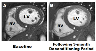
Cardiac MR with forced detraining of the athlete is a useful strategy to differentiate between athlete's heart and HCM. Patients who decondition over a 3-month period, and in whom wall thickness regresses greater than 2 mm, supports a diagnosis of Athlete's Heart while hypertrophy that remains unchanged suggests HCM. 


A. Baseline Cardiac MR. 
B. Repeat CMR following 3-month deconditioning showed no change in LV wall thickness. This suggests a diagnosis of HCM. 


-YZ-

Reference:

Can too much exercise be dangerous: what can we learn from the athlete’s heart? (bjcardio.co.uk)

Athlete's heart or hypertrophic cardiomyopathy? - PubMed (nih.gov)

Distinguishing hypertrophic cardiomyopathy from athlete’s heart physiological remodelling: clinical significance, diagnostic strategies and implications for preparticipation screening | British Journal of Sports Medicine (bmj.com)

Cardiovascular Magnetic Resonance Imaging in the Assessment of Athletes with Heart Disease - American College of Cardiology (acc.org)

Complications of Group A Strep Infection

Complications of Group A Strep Infection

Group A strep also known as Streptococcus pyogenes is a common gram positive cocci that can cause a wide range of infections in humans. Common infections include a pharyngitis ("strep throat") and skin and soft tissue infections. Here we will review the common presentations of Group A strep infections and their complications. 

Infections Presentations

Infections of the Head and Neck

- Pharyngitis

- Tonsillitis

- Peritonsillar Access 

- Otitis Media

Skin and Soft Tissue Infections

- Erysipelas 

- Cellulitis

- Impetigo

- Necrotizing Fasciitis

- Perianal streptococcal dermatitis

Non-Infections Presentations

One of the virulence factors of Streptococcus pyogenes is through toxins. The three main toxins that strep pyogenes produces are: 1) Erythrogenic exotoxin A 2) Streptolysin O and 3) Streptococcal pyrogenic exototoxins

Toxin-mediated diseases

- Scarlet Fever

    - The erythrogenic exotoxin A, B or C produced by strep pyogenes can be transmitted via aerosols and cause a delayed-type skin reaction. The clinical features of scarlet fever include a scarlet-coloured maculopapular rash which is fine, erythematous and sandpaper-like in texture, flushed cheeks, perioral pallor, strawberry tongue. 7-10 after resolution of rash desquamation of skin in flakes may occur. 

- Streptococcal toxic shock-like syndrome

    -While toxic shock syndrome is caused by Staph aureus, strep pyogenes can also produce a toxic shock-like syndrome which is caused by release of its pyrogenic exotoxin. Clinical features include: Fever, flu-like symptoms, transient erythematous macular rash, strawberry tongue, conjunctivitis, and symptoms of shock if severe. 

Immunologic Complications 

- Acute Rheumatic Fever 

    - Acute rheumatic fever (ARF) is an inflammatory sequela involving the heart (pancarditis), joints (migratory polyarthritis), skin (subcutaneous nodules and erythema migrans) and CNS (Sydenham Chorea) that occurs two to four weeks after an untreated infection with GAS infection

- Poststreptococcal Glomerulonephritis

    -An acute glomerular inflammation that results from a preceding infection with nephritogenic strains of group A (beta-hemolytic) Streptococcus pyogenes. Most commonly seen in children approximately 10–30 days following group A streptococcal tonsillopharyngitis or possibly skin infections such as impetigo.

- Acute Post-streptococcal polymyalgia

Acute Post-streptococcal polymyalgia is a rare immunological phenomenon of a GAS infection. Common features include sudden onset of muscular pain with fever usually after an acute upper respiratory tract infection. 

References: 

Rosenthal KS, Tan MJRapid Review Microbiology and Immunology, 3rd EditionElsevier; 2010

Venuta A, Ferrari P, Beverelli A, Bettelli F, Laudizi L. Acute post-streptococcal polymyalgia: two new cases with a review of the literature. Eur J Pediatr. 1995 Jun;154(6):475-6. doi: 10.1007/BF02029359. PMID: 7671947.

-AM-


An Approach to Heart Failure and Pulmonary Edema on Chest X-ray

 An Approach to Heart Failure and Pulmonary Edema on Chest X-ray

It is important to note that pulmonary edema in the lungs follows, a certain pattern of progression, which will show up on imaging. Understanding of these imaging findings requires first and understanding of heart pressures and an underlying pathophysiology of heart failure.

One of the most important underlying features of left sided heart failure is elevated left-sided heart pressures. In many cases with impaired systolic function, the left ventricle is unable to eject blood forward and therefore there will be elevated chamber pressures in both the left ventricle and left atrium. After a prolonged period of time, there will be a retrograde transmission of elevated pressure into the pulmonary veins (which are directly connected to the left atrium). If the pressures are high enough, it will cause transudation of fluid outside of the pulmonary veins into the interstitium surrounding the alveoli, and eventually transudation of fluid directly into the air spaces. 


These sequential steps in left sided heart failure also produce characteristic imaging findings which corresponds to the stage of pulmonary edema.

These three distinct stages are:

1.)  Pulmonary venous congestion

2.)  Pulmonary interstitial edema.

3.)  Pulmonary alveolar edema

While it is difficult to directly measure, left atrial pressure, there is a surrogate for this, and it is pulmonary capillary, wedge pressure (PCWP). Is Sepand in the catheter lab where a catheter is advanced into the right atrium than the right ventricle and advanced all the way maximally to the pulmonary capillaries. The pressure reading at the pulmonary capillaries is a direct measure of left atrial pressure. A normal PCWP is between 8-12mmHg, meaning a normal left atrial pressure is also between 8-12 mmHg. 

Pulmonary Venus congestion occurs at a PCWP of 13 - 18 mmHg, pulmonary interstitial oedema occurs at a PCWP of 18 - 25 mmHg, and finally alveolar edema occurs at PCWP of > 25 mmHg. 

Imaging Findings

Pulmonary Venous Congestion

This is the first stage of heart failure, and imaging findings can be very subtle. In the lungs, the pulmonary vessel supplying the upper lung fields are smaller and fewer number, and those at the long bases. This can be identified as the upper lung fields are often more lucent and darker than the lower lung fields.

In the following imaging to be clearly seen that there are higher calibre than normal pulmonary vans in the upper lung fields. There are not yet any findings of interstitial or alveolar pulmonary oedema and this represents an early sign of heart failure. This may be also called civilization, vascular, redistribution, or stags antlers sign.

Case courtesy of Calum Worsley, Radiopaedia.org, rID: 91249

The following picture from the website, Radiology Assistant also demonstrates a picture of a patient in good condition on the left, and during a period of CHF, where we can clearly see increase vascular predominant in the upper lung fields, again representing vascular, distribution or cephalization. This is an example of pulmonary Venous congestion.


Pulmonary Interstitial Edema

In pulmonary interstitial edema, the pressures inside the venous system are elevated enough that fluid translates across the endothelial lining of pulmonary veins into the tissue between the vessels and the alveoli this gives the characteristic wet appearance in the lungs and occurs at left atrial pressures between 18 to 25 mmHg. 

The radiographic features of pulmonary interstitial oedema include Peribronchial cuffing, Kerley B lines, and thickening of the interlobar fissures. 

In the following image, not only can vascular redistribution be seen, but also features of pulmonary interstitial edema, including Kerley B lines, thickening of interlobar fissures, and cardiomegaly.

Case courtesy of Abeer Ahmed Alhelali, Radiopaedia.org, rID: 51448


Pulmonary Alveolar Edema

The final stage of pulmonary oedema is alveolar edema. In this stage fluid continues to leak out of the vessels, into the interstitium, and into the air spaces themselves. This occurs at left atrial pressure is greater than 25 mmHg.

Radiographic features of pulmonary alveolar edema include opacities in the lung fields that are fan-shaped outwards from the hilum in a "batwing" pattern. The opacities may look like dispersed cotton balls in the lung – this is very characteristic of pulmonary alveolar edema finally air bronchograms, pleural effusions, and cardiomegaly are often also seen in this stage of pulmonary edema.


In conclusion, acutely decompensated heart failure can have different patterns in the lungs and correspond to the progression of disease, which in order are: pulmonary venous congestion, pulmonary interstitial edema, and pulmonary alveolar edema.

References:


-AM-